Posts

Neoplasia

Image
      Neoplasm (Cancer)           Neoplasm is new tissue growth that is unregulated, irreversible and monoclonal . Monoclonal means the cells are derived from a single mother cell. Similarly, the term 'hyperplasia' is definied as the cells are not derived from a single parent cell. Moreover hyperplasia may be pathological and some times physiological. Hyperplasia is reversible to a certain period. Hence Hyperplasia is not consider as cancer.           Cancer is the 2nd leading cause of death. Most common caners which incidence in adults are breast/prostate cancer, lung cancer, and colorectal cancer (cancer in large intestine). Causes of neoplasm:           Carcinogens are the substance which increase the DNA damage which develops to cancer.                                              Chemical carcinogens include:                                                      1. Alcohol                                                      2. Arsenic                                    

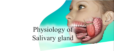
Physiology of Salivary gland

Image
Physiology of Salivary gland Saliva is a biological fluid which performs functions like bolus formation, secretion of enzymes, excretion of chemical substances etc. We have 3 pairs of the major salivary gland and some minor salivary glands. Major salivary glands perform maximum involvement in salivary secretion. Major salivary glands: Major salivary glands include 1.      Parotid glands 2.      Submandibular glands 3.      Sublingual glands On both side of our buccal cavity. 3 major salivary gland Parotid glands: They are the largest of all salivary glands. It is located on the lateral surface of oral cavity. This gland communicates to the oral cavity by Stenson's duct in the upper 2 nd molar tooth. Submandibular glands: These glands located in the submaxillary triangle and communicates through Wharton’s duct. It opens at the frenulum of the tongue by an opening called Caruncula sublingualis. Sublingual glands: The Smallest salivary gland located on t

Humoral & Cell mediated immunity

Image
     Our immune response is done by specialized blood cells in our body combined with immune organs. Based on the type of response expressed by the immune cells, the human immune response is of two types, humoral immunity and cell mediated immunity.      Humoral immunity immune response is mostly involved B lymphocytes (B cells) and cell mediated immune response only involve T lymphocytes (T cells). Immune cell formation in short:      From the hematopoietic cells in bone marrow, precursor cells of B and T cells are formed. B cells undergo maturation in the blood. Whereas T precursor cells reach the thymus gland near to the heart. From the thymus, T precursor cells differentiate to T helper cells (CD4) and T killer cells (CD8). Short form of immune cell production Humoral immune response:     When an antigen enters our blood system, It will engulf by antigen presenting cells (APC), especially macrophages. Then it will undergo a process and produce antigen peptide. All the APCs have MHC

Inflammation

Image
Inflammation:      It is a physiological process that is expressed by our immune system and may convert to pathological conditions in the future based on the severity of the inflammation.      It is classified into Acute and chronic inflammation based on the components of the inflammatory process. Inflammatory mediators: These are the chemical compounds that initiate and maintain the inflammatory process. 1. Arachidonic acid metabolites:      Arachidonic acid is released from the cell membrane by phospholipase A2. Then it will react with cyclooxygenase or 5-lipoxygenase enzymes.          - Arachidonic acid with cyclooxygenase produces prostaglandins (PG), which mediate vasodilation.          - Arachidonic acid with 5-lipoxygenase produces leukotrienes (LT), which mediates vasoconstriction and bronchospasm. 2. Mast cells: Mast cells are activated by any trauma and by complement proteins C3a and C5a. Mast cells act 2 forms of response. Immediate and delayed response.          - Immediate

Jaundice

Image
Introduction:      Jaundice is a pathological process of bilirubin metabolism. Bilirubin a catabolic product of the "heme" part of hemoglobin. Naturally, bilirubin is yellow in color. That's why our body color change to yellower in jaundice. Usually occur >2.5 mg/dL of bilirubin in serum. Normal bilirubin metabolism:      RBCs are consumed by macrophages after their life span (120 days) by the reticuloendothelial system. During this process, hemoglobin present in RBCs will get disintegrated to heme+globin. From the heme part, protoporphyrin is converted to unconjugated bilirubin (UCB). Albumin will take this UCB to the liver. In the liver, we have an enzyme called Uridine Glucuronly Transferase (UGT) in the hepatocyte. This enzyme will convert the UCB to conjugated bilirubin (CB).      This CB will transfer to the bile canal and forms the bile, which will store in the gall bladder. When the digestion in the small intestine begins, the bile will be released from the gal

Pathophysiology of COVID 19

Image
Introduction:                                           COVID 19 is a viral respiratory disease that is able to cause systemic inflammatory response syndrome from the lungs. This virus was originated from China and it can spread through cough and fecal matter from an infected person. In this blog, I explain the clear pathophysiology of this disease.       Basically, COVID 19 is caused by Coronavirus. Microbiologically it is a positive single-strand RNA virus (+ ss RNA). It is a complex virus which means it has a capsid inside the envelope. As the name indicates, Coronavirus have spikes in the envelope. It is the largest RNA virus in which the genome is approximately 30 kb.      Coronavirus have nucleoprotein (capsid) around the RNA which is called N protein. The lipid bilayer membrane is covered as an envelope (E protein). The envelope has glycoprotein spikes on them called S protein. Etiology (Causes):      COVID 19 is spread mostly through airborne and from fecal matter. It is believ

Consciousness & Stages

Image
CONSCIOUSNESS Mainly two types of consciousness. 1.  Clear  consciousness (Normal state) 2.  Affected consciousness We have 4 stages of affected/Altered consciousness. 1. Clouding of conscious 2. Stupor 3. Sopor 4. Coma Let discuss briefly in this blog!