Infectious endocarditis (IE)
Introduction:
Infective endocarditis (IE) is a microbial infection of a heart valve (native or prosthetic) or the endocardium, leading to tissue destruction and formation of vegetation. Caused mainly due to streptococcal and staphylococcal infection in the heart valves.
Etiological statistics:

Development of Aschoff nodules in IE:
The cardiac manifestations are in the form of focal inflammatory involvement of the interstitial tissue of all the three layers of the heart, the so-called pancarditis. The feature of pancarditis in RF is the presence of distinctive Aschoff nodules or Aschoff bodies. The Aschoff nodules are spheroidal or fusiform distinct tiny structures, 1-2 mm in size.

Fully-developed Aschoff bodies occurs through 3 stages:
1. Early (exudative or degenerative)
stage
2. Intermediate (proliferative or granulomatous) stage
3. Late (healing or fibrous) stage
Early (exudative or degenerative) stage:
üThe earliest sign of injury in the heart is about 4th week of illness.
üEdema of the connective tissue and increase in acid mucopolysaccharide in the ground substance.

Intermediate stage:
üThis stage is apparent in the 4th to 13th week of illness.
üThe early stage of fibrinoid change is followed by proliferation of cells that includes infiltration by lymphocytes (mostly T cells), plasma cells, a few neutrophils and the characteristic cardiac histiocytes (Anitschkow cells) at the margin of the lesion.
üThese cardiac histiocytes become multinucleate cells containing 1 to 4 nuclei and are called Aschoff cells.
Late (healing or fibrous) stage:
üThe stage of healing occurs about 12 to 16 weeks after the illness.
üThe nodule becomes oval or fusiform in shape, about 200 µm wide and 600 µm long.
These cells tend to be arranged in a palisaded manner.

IE develops in heart, in a manner of layer by layer associated with rheumatic heart disease (RHD).
1. Rheumatic endocarditis- Rheumatic valvulitis
2. Rheumatic myocarditis
3. Rheumatic pericarditis
But for a look, rheumatic endocarditis is the only pathologic condition that associate with infectious endocarditis.
Development of IE Associated with Rheumatic endocarditis:

The approximate frequency of deformity of various valves:
According to the data, the mitral valve is almost always involved in RHD and IE.
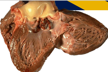
Gross appearance of chronic healed mitral valve in RHD and IE is characteristically ‘fish mouth’ or ‘buttonhole’ stenosis.
Diagnosis of IE:
üClinical featuresüMicrobiological analysis
Microbiological analysis:
Blood cultures are positive in about 80% of cases, but maybe negative in cases of intracellular or fastidious pathogens or after previous antibiotic treatment.
Echocardiography:
üA transthoracic echocardiogram (TTE) should be performed promptly.
üTransoesophageal echocardiography (TOE) offers better image quality and the overall sensitivity for IE is 90–100%.
Three echocardiographic findings are the major criteria for the diagnosis of IE.
(ii) Peri annular abscess;
(iii) new dehiscence of a valvular prosthesis.
Vegetation size and mobility is important.
If the vegetations are small or have already embolized, echocardiography can produce false-negative results in about 15% of cases.
Treatment for IE:

Surgical Importance:
Antimicrobial therapy can offer a curative treatment in only 50% of patients.
The surgical aim is valve repair, but most patients require valve replacement. Patients with IE and large vegetations, intracardiac abscess (9–14%), or persisting infection (9–11%) almost always need surge. The current IE perioperative mortality is 5–15%. The most frequent postoperative complications are persistent septic shock, coagulopathy, acute renal failure, stroke, refractory heart failure, and conduction abnormalities.
Conclusion:
Epidemiology of IE makes the diagnosis a challenge, and traditional diagnostic criteria are insufficient. Despite modern medical and surgical therapy, IE is still associated with a high rate of complications and increased mortality. Early surgery is becoming more common and TOE should be used for all patients.


Comments
Post a Comment